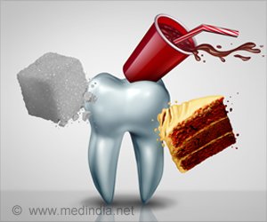
Restorative materials do not always bond well to the surrounding healthy tooth structure. Microscopic leaks may form, allowing fluids and bacterial acids to penetrate beneath the restoration. This can lead to the formation of a secondary caries that appears and grows around a previously restored cavity.
Dentists now spend more time replacing failed restorations than placing new ones due to the maladaptation of bonding materials to tooth structure.
Developing New Diagnostic techniques to Detect Active Dental Lesions
In a recent study published in Journal of Biomedical Optics (JBO), a research team evaluated emerging imaging modalities for discerning active tooth decay.
Advertisement
The traditional methods relying on tactile sensation via a dental explorer and visual inspection based on texture and color are highly subjective and unreliable. There is also no established imaging technology that can provide information with high specificity and sensitivity when assessing dental decay activity.
To address this issue, the researchers focused on whether shortwave-infrared (SWIR) and thermal imaging could be combined with air drying to accurately diagnose the activity of a secondary caries lesion.
The idea underlying both these methods is that active lesions are more porous than healthy teeth, and these pores hold water.In the SWIR-based approach, one can indirectly detect active lesions by observing changes in the SWIR reflectivity as the tooth dries out.
On the other hand, the thermal imaging-based approach relies on the fact that the temperature changes in active lesions during air drying are different from that in healthy teeth, owing to the water trapped in the pores of the lesion.
In their work, the team acquired 63 human tooth samples from oral surgeons and analyzed 109 suspected secondary lesions in them using both SWIR and thermal imaging.
In addition to these methods, they also observed the samples using optical coherence tomography (OCT), a more sophisticated technique that uses near-infrared light to create high resolution 3D images.
To determine whether SWIR and thermal imaging were indeed useful for detecting active lesions, the results of these methods were compared with those obtained via OCT. Overall, SWIR proved superior to thermal imaging and performed better in most circumstances.
The SWIR permeability measurements were well correlated with the thickness of the transparent surface layer (TSL) of lesions measured via OCT. They also found that the highly mineralized TSL was thickest when a lesion had been fully arrested and needed no further intervention.
According to the OCT results, a TSL thicker than 70 µm was a potential indication that a lesion was no longer active. The findings of this study could help pave the way to a new era of diagnostic imaging in dentistry.
Source: Eurekalert
Source link
#Detect #Tooth #Decay #Accurately #Invisible #Light