‘Stiffness’ of cells could give early warning of cancer

A new technology developed in the UK could make it possible to detect cancer at an early stage, long before the formation of a lump, by investigating the stiffness of individual cells.
Researchers in the UK have developed an endoscopic device that uses 3D imaging to look at the stiffness or ‘viscoelastic’ properties of individual cells in situ, without the need for a biopsy or the use of markers like fluorescent tags or contrast media.
The team – from the University of Nottingham – notes that in the early stages of their development cancer cells are much softer than healthy cells, a property that helps them squeeze through gaps and spread locally and to distant parts of the body.
As they spread, the malignant cells also modify their surrounding environment to create an abnormally stiff 3D extracellular matrix of macromolecules that protects tumours as they develop.
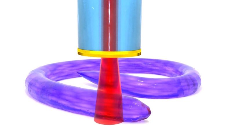
The new technology – billed as a world-first by its developers – uses a physical phenomenon called Brillouin scattering to measure the stiffness of individual cells with a hair-thin endoscopic probe. It opens the door to investigating microscopic cellular tissue based on abnormal stiffness at the single-cell level inside the human body for the first time.
“Our device makes it possible to ‘feel for a stiff lump,’ but on a single cellular scale, meaning we could catch cancer early at microscopic cell scales rather than large malignant tumour scale,” said lead author Dr Salvatore La Cavera III, a research fellow in Nottingham University’s optics and photonics group.
“We aim to develop new endoscopic technologies that make diagnostics faster, safer, and clearer for both patients and clinicians,” he added, without the discomfort of a biopsy and the logistical challenges of processing, transporting, and analysing samples.
So far, the researchers have tested the capabilities of the technology using a living nematode worm as a model. In a paper published in the journal Nature Communications Biology, they show the device was able to give a reading of the biomechanical properties of the cuticle, a collagen-rich structure that protects the worm from its environment and is also used as a lab model for biological processes in other species.
Until now, it has only been possible to image the cuticle using electron microscopes in non-living conditions, but the device was able to reveal the physical surface properties of a fully formed microscopic organism “in unprecedented detail,” according to co-author Dr Veeren Chauhan of the university’s school of pharmacy.
“This capability will not only enhance our understanding of nematode biology, but also builds a foundational model for translating these findings to complex biological systems, including human physiology, paving the way for advancements in both basic biological research and clinical diagnostics,” he added.
Source link
#Stiffness #cells #give #early #warning #cancer